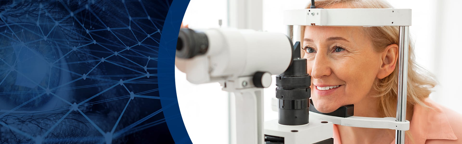
Your Eye Health Comes First
Your long-term eye health is a priority for our team at Great Lakes Optometry. Eye disease can come in many different forms and jeopardize your eye health in many cases. Our team wants to help preserve your long-term vision, whether you’re dealing with cataracts, glaucoma, age-related macular degeneration, or diabetic eye disease.
We recommend consistent eye exams so that our team can address any issues with your eyes early. Our approach to detecting, monitoring, and managing eye disease is hands-on and thorough.
Contact us to schedule your appointment today and get a head start on eye disease management.
Book AppointmentEarly Detection Through Technology
The earlier we detect signs of eye disease, the sooner our team can take action and begin preparing your personalized treatment plan to preserve vision.
We implement diagnostic technology such as OCT, visual field testing, tonometry, and more to capture the details of your eye health and get started on a personalized treatment plan early to preserve your long-term vision.
The Different Forms of Eye Disease
Patient education is important to us here at Great Lakes Optometry. Eye diseases can affect your eye health and vision in many ways.
By sharing the different forms of eye disease and making you aware of some of the early warning signs, we can help you understand your risks for certain conditions and highlight the importance of regular eye exams.
Our team works to address potential eye disease issues early before your vision and eye health are affected.
Glaucoma
Glaucoma is a group of eye conditions that cause damage to your optic nerve, which is responsible for sending visual information to your brain. Damage to your optic nerve can lead to vision loss and blindness if not detected and treated early.
Damage from glaucoma can be caused by high pressure in the eye, referred to as intraocular pressure. Age, family history, and previous eye injuries can all be glaucoma risk factors. Maintaining a regular eye exam schedule is essential for detecting glaucoma early.
We check for glaucoma in every single patient and offer tonometry, which is an eye test that can detect changes in eye pressure long before you may be aware of them.
Cataracts
Cataracts develop when the normally clear lens of the eye becomes clouded and can cause symptoms like blurry vision, dim or yellowed vision, and difficulty seeing at night. Cataracts begin developing when proteins in your eye form clumps that partially obscure your lens and stop light from reaching your retina, the thin layer of tissue that lines the back of your eye.
If cataracts progress to a later stage and prevent you from going about your daily activities, such as reading or driving, cataract surgery is an option to remove them.
We offer pre-cataract surgery evaluation and post-op care.
Age-Related Macular Degeneration
Age-related macular degeneration (AMD) is a progressive disease that affects your central vision, damaging your macula. This damage can make things like reading and driving much more difficult.
AMD is a leading cause of vision loss among older adults, and there are 2 main types:
-
- Dry AMD is the more common form of the disease and occurs in early, intermediate, and late stages. This form of AMD occurs when the macula thins with age.
-
- Wet AMD is the less common form of the disease and can cause faster and more severe vision loss. Wet AMD occurs when abnormal blood vessels grow in the back of the eye and damage the macula.
Our exams include OCT and visual field testing to help detect and manage AMD.
Diabetic Eye Disease
Diabetes can increase your risk of developing eye diseases such as glaucoma, cataracts, and diabetic retinopathy, which is a primary vision loss concern for patients with diabetes.
Diabetic retinopathy is a condition that causes damage to the blood vessels of the retina. Symptoms of diabetic retinopathy can include:
-
- Black spots or holes in your vision
-
- A loss of central vision
-
- Blurry vision
-
- Trouble seeing at night
We use optical coherence tomography (OCT) and retinal imaging in our diabetic eye exams to assist with the early detection of diabetic eye issues.
Amblyopia (Lazy Eye)
Amblyopia, also known as lazy eye, is the loss or lack of development of clear vision in 1 or both eyes. Amblyopia typically develops at a younger age, and eyeglasses or contact lenses may not be able to fully correct the reduced vision caused by amblyopia.
Some causes and risk factors can include:
-
- Family history
- A high prescription that’s gone uncorrected with glasses or contacts
- Eye turn
- Large refractive errors
- Visual deprivation of one eye
Treatment for amblyopia can include a combination of prescription lenses, prisms, vision therapy, and more. Our team will work with you to find the right treatment for your needs.
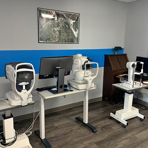
Our Diagnostic Technology
Our clinic is equipped with modern technology and different imaging equipment to help diagnose and manage common eye diseases such as glaucoma, diabetic retinopathy, cataracts, and AMD.
Our diagnostic technology includes OCT, visual field testing, and more to help our team detect signs of eye disease early. Early detection allows our team to get a head start on preparing a personalized treatment method for your needs.
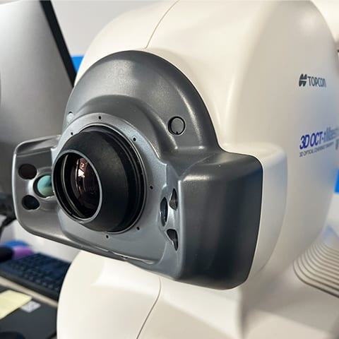
Optical Coherence Tomography
We use optical coherence tomography (OCT) to help us monitor retinal conditions that can lead to profound vision loss or blindness. These common conditions include diabetes, high blood pressure, glaucoma, and macular degeneration.
OCT is a noninvasive imaging technique that uses reflected light to image the back of your eye, including the retina and optic nerve.
Visual Field Testing
Visual field testing is used to help us see what you see (and what you do not see). It has been a gold standard for the diagnosis and management of glaucoma for decades. People with glaucoma typically need a threshold visual field test performed at least once a year.
As the eye is an extension of the brain, it is also possible to diagnose certain brain lesions with visual field testing.
The visual field testing helps our team visualize what advanced glaucoma can look like.
Tonometry
We offer tonometry to help detect elevated intraocular pressure (IOP) early because of glaucoma. Our practice features the iCare tonometer and Goldmann tonometry.
The iCare tonometer is a diagnostic tool used during our eye exams to measure IOP. Unlike other traditional tonometry methods, the iCare tonometer offers quick and easy IOP measurements without the need for anesthesia or puffs of air.
Fundus Photography
Fundus photography is a noninvasive diagnostic tool that captures detailed images of the back of your eye, allowing our team to detect and monitor various eye conditions.
With fundus photography, we can betteGlaucoma is a group of eye conditions that cause damage to your optic nerve, which is responsible for sending visual information to your brain. Damage to your optic nerve can lead to vision loss and blindness if not detected and treated early.
Damage from glaucoma can be caused by high pressure in the eye, referred to as intraocular pressure. Age, family history, and previous eye injuries can all be glaucoma risk factors. Maintaining a regular eye exam schedule is essential for detecting glaucoma early.
We check for glaucoma in every single patient and offer tonometry, which is an eye test that can detect changes in eye pressure long before you may be aware of them.
Monitoring Your Visual Health
Our philosophy when it comes to diagnosing and managing eye disease is to practice early detection and management. We encourage our patients to schedule regular, consistent eye exams so we can get a head start on any potential issues.
Don’t wait for your vision to be affected—contact us to schedule your appointment today.
Book AppointmentOur Location
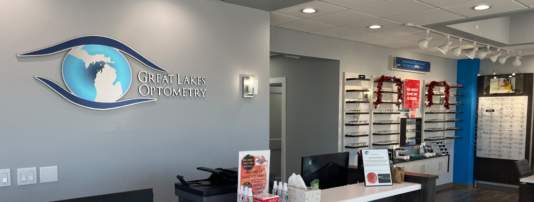
Our Address
- 2237 Ashmun Street
- Sault Ste. Marie, MI 49783
Contact Information
- Phone: 906-635-9600
- Fax: 906-635-1077
- Email: [email protected]
Our Hours
Hours
- Monday: 8:00 AM – 5:00 PM
- Tuesday: 8:00 AM – 5:00 PM
- Wednesday: 8:00 AM – 5:00 PM
- Thursday: 8:00 AM – 5:00 PM
- Friday: 8:00 AM – 4:00 PM
- Saturday: Closed
- Sunday: Closed
Our Brands


Our Google Reviews
-
Great Lakes Optometry
- 2237 Ashmun Street
- Sault Ste. Marie, MI 49783
-
P: 906-635-9600
[email protected]